Breaking

Monday, July 23, 2018

7/23/2018 04:43:00 PM

stroke

Introduction
Stroke has cerebral infarction as a disease in which blood vessels are blocked and intracranial hemorrhage as a disease that blood vessels break (Figure 1)
stroke
Here, we will focus on representative diseases that are treated by neurosurgery, among various strokes.

subarachnoid hemorrhage
Subarachnoid hemorrhage is characterized by a sudden headache. It is expressed as strong pain that I have never experienced before, "pain like beat with a bat" or "pain like lightning thunder". Others may cause vomiting and disturbance of consciousness. Very light ones may have mild headache, dizziness, nausea only. It happens for various reasons (Figure 2),
subarachnoid hemorrhage
The most common thing is the rupture of a Cobb (cerebral aneurysm) formed in a blood vessel of the brain. CT diagnosis is useful for diagnosis (Figure 3).
subarachnoid hemorrhage
In light bleeding, diagnosis with CT is sometimes difficult. In that case, MRI examination and cerebrospinal fluid examination are used together. Because the death rate due to reruption of aneurysm is very high, it is necessary to have a quick diagnosis and treatment in a specialized facility. If a cerebral aneurysm that causes bleeding is found, intravascular surgery will be performed such as clipping an aneurysm by surgery or filling aneurysm with a special coil (Fig. 4).
Treatment of cerebral aneurysms
In 2011, Kumamoto Prefecture's subarachnoid hemorrhage survey, 383 people in all prefectures underwent subarachnoid hemorrhage in one year, and 20.9 people occurred per 100 thousand population. From 60s to 80s average age was 67.1 years old. About 50% of those who had good treatment outcome, about 70% among those who could be treated (Kumamoto Prefecture Cerebrovascular Injury Data Bank). It is a very scary disease that requires immediate medical treatment and delays in diagnosis and treatment may cause severe sequelae. In case of sudden severe headache or unusual headache, please get medical examination as soon as possible.

cerebral hemorrhage
Cerebral haemorrhage is often referred to as hypertensive cerebral hemorrhage because it occurs mainly due to high blood pressure after the middle age. Recently, there have been cases of cerebral hemorrhage occurring due to other drugs such as warfarin and aspirin because they take medicine that makes blood hard to clump. Cerebral haemorrhages such as children and young people who do not have a history of hypertension are likely to be frequently bleeding due to blood vessel malformations.
Depending on the site of bleeding, it is divided from putamen bleeding, thalamic bleeding, cerebral hemorrhage, bridge hemorrhage, subcortical bleeding, etc. (Figure 5).
cerebral hemorrhage
Symptoms vary according to the site of bleeding, but hemiplegia, aphasia, disturbance of consciousness, etc. are recognized. It is diagnosed by CT and MRI and angiography are performed as necessary. For treatment, systemic management and blood pressure management are strictly performed first, and operation indication is decided taking into consideration symptoms, life prognosis, function prognosis, age, hematoma volume, etc. Surgery includes removal of craniotomas, stereotactic or endoscopic hematoma aspiration (Figure 7).
cerebral hemorrhage
cerebral infarction
Among cerebral infarctions, lacunar infarction caused by clogging of thin blood vessels in the brain and atherothrombotic cerebral infarction that occurs due to clogging of the thick artery of the brain with thrombus caused by arteriosclerosis, thrombus formed by the heart obstruct the artery of the brain There is a cardiogenic cerebral embolism that occurs to do. In general, medical treatment is the main subject. Among them, "carotid artery stenosis" is said to occur at the origin of the internal carotid artery in the cervix, atherosclerosis occurs, diseases causing cerebral ischemic attacks and infarction due to cerebral blood flow reduction and thrombus formation as an embolus source Yes. As a treatment, a surgical operation (carotid endarterectomy) to remove thickened atherosclerotic intima (FIG. 8)
cerebral infarction
Endovascular surgery, drug treatment and so on. We also know that treatment results of endarterectomy are better than medications when the stenosis is strong. In addition, due to internal carotid artery obstruction and blood flow decrease due to stenosis and occlusion of intracranial vessels, bypass surgery may be performed in cases of cerebral infarction and transient ischemic attacks.

Endovascular operation
Endovascular surgery is a treatment method using a catheter, but it is a field that has developed remarkably due to recent improvements in catheters and embolic materials, advances in imaging equipment, technological improvement of catheter operation by surgeons, and so on. It is useful not only for treatment of aneurysm, but also for treatment of cerebral vascular malformations, dural arteriovenous malformations and so on. It is also becoming possible to inflate thinned blood vessels with a balloon (balloon), or treat a stent by expanding it from the inside to the stenotic lesion of the carotid artery (Fig. 9).
Endovascular operation
It seems that it will develop further in the future, and many patients are expected to benefit from this.

Finally
The above is the explanation focusing on treatment to be performed by neurosurgery in stroke. However, prevention of stroke is more important than anything else. Hypertension, diabetes, hyperlipidemia, atrial fibrillation, smoking etc. are cited as risk factors for stroke, but please consult with your family teacher to receive these appropriate treatments. Also, let's take into consideration diet, exercise, weight control, alcohol intake and so on.
If there is something to worry about, please do not hesitate to consult us.
7/23/2018 04:42:00 PM

Diet and diabetes

concept
Diabetes is characterized by chronic hyperglycemia due to lack of insulin action and is a group of diseases accompanying various metabolic abnormalities. Due to persistence of long-term hyperglycemia etc., complications specific to diabetes (three major complications (diabetic retinopathy, nephropathy, neuropathy, etc.), etc.), arteriosclerosis and other complications may be caused. Currently it is increasing in Japan, with about 8.9 million people suspected strongly of diabetes and 22 million people estimated to be combined with those who can not deny diabetes. There are about 3,000 people (2nd place of cause of blindness in adults) in one year due to diabetes, about 16,000 people introduce dialysis (first place of dialysis cause), hands and feet It is estimated that there are several thousand people to cut. Furthermore, it is known that cerebral infarction and myocardial infarction are also liable to occur threefold when diabetes is present, and the course after onset is also bad.


Symptom
Symptoms such as dry mouth, polydipsia, polyuria, and weight loss appear if it gets worse, but there are also cases where there is no subjective symptom. Sometimes it is diagnosed as diabetic by visiting a hospital with symptoms due to complications (eg abnormal appearance).

Cause
In addition to inheritance (the constitution born with it), type 2 diabetes which develops with lifestyle affects accounts for nearly 90%. In addition to this, type 1 diabetes, in which insulin secretion is rapidly depleted due to autoimmune etc., diabetes that develops with other diseases, diabetes related to pregnancy, and so on.

Inspection for diagnosis
The normal value of blood glucose is less than 110 mg / dl on an empty stomach. Diabetes is strongly suspected in cases of fasting blood glucose level of 126 mg / dl or more, blood glucose level as occasionally (regardless of meals) 200 mg / dl or more, HbA 1 c 6.5% or more, It may be diagnosed by 75 g oral glucose tolerance test.

Treatment
There are life therapy (diet therapy, exercise therapy, smoking cessation, drinking to keep an appropriate amount, etc.) and medication therapy (internal medicine, injection drug (insulin preparation, GLP-1 analog preparation)).

prevention
Diet therapy is the foundation of prevention and treatment of diabetes. Although it is basic to not eat too much over the appropriate amount (optimization of total calories), it is also important to ingest the necessary nutrients balancedly as daily nutrition. Generally avoid excess of salt and alcohol, vegetables and dietary fiber intends to ingest enough.
Recently "saccharide restricted diet" may become a topic. A good effect can be expected for those who eat too much every day, such as "to refrain from sweet things" "to moderate rice". However, diet therapy that restricts carbohydrates (carbohydrates) extremely and supplements calories with other (fat and protein) is currently not recommended by the Japanese Diabetes Society from the aspect of safety. Compared to the 1960s, when diabetes was still few in Japan, the intake of total calories and carbohydrates gradually decreased, while the intake of fat has increased greatly. At that time, it seems that the intake of rice per capita was about twice as much as the current amount and the intake of fat was about half. It seems that the Japanese staple food has changed from rice to bread, men and others, and has begun to like butter, lipids, dairy products (so-called Western-style meals). At that time, it should be noted that the intake of dietary fiber such as millet, root vegetable and so forth was larger (about 1.4 times) than the present amount (thought to be) (thought to be) (thought to be) a large amount of physical activity, but the investigation of Japanese who moved to Western Europe It is also known that diabetes is seen at a high rate. There seems to be a hint of "diet for prevention of diabetes" among these.
7/23/2018 04:39:00 PM

Obesity and metabolic syndrome

concept
Metabolic syndrome is a condition in which arteriosclerotic diseases such as visceral obesity, insulin resistance / hyperglycemia, abnormal lipid metabolism, rise in blood pressure and the risk of developing type 2 diabetes are accumulated personally. Even if it seemingly does not seem to be an illness or abnormality slightly exceeding the normal value, if it accumulates in an individual, the above risk factor (the risk of cardiovascular disease onset and cardiovascular death is 1.5 to 2 Twice, the risk of developing type 2 diabetes increases 3 to 6 times). Originally it has been proposed as a risk factor other than LDL (bad) cholesterol known as a risk factor for coronary artery disease, so LDL cholesterol is not included in the diagnostic criteria. Hypertension, diabetes, dyslipidemia and obesity are also risk factors for atherosclerotic diseases (eg, about 3 times for diabetes and about 2 times for borderline diabetes mellitus), and these are "slightly abnormal" If you are staying in the treatment area, you need to take more seriously and deal with it.


Symptom
Abdominal girth, blood pressure, etc. can be measured by self, but there is generally no subjective symptom.

Cause
Obesity (especially visceral obesity) is thought to play a central role. Adipocytes are known not only to store fat but also to emit various bad substance. It is also known that obesity not only metabolic syndrome but also many diseases merge.

Inspection for diagnosis
The waist perimeter (abdominal circumference) is 85 cm or more in men and 90 cm or more in females, and it is diagnosed as metabolic syndrome if it falls into two or more of the following three items.

Neutral fat (fasting) 150 mg / dl or more and / or
HDL cholesterol less than 40 mg / dl
Systolic blood pressure of 130 mmHg or more and / or diastolic blood pressure of 85 mmHg or more
Fasting blood glucose 110 mg / dl or more
(In case of drug being treated already, treat it even if it is below the reference value)
Treatment and Prevention
As for metabolic syndrome, obesity plays a central role, so it is important to maintain proper weight in diet therapy, exercise therapy and other life therapy. In addition, smoking cessation is extremely important in preventing arteriosclerosis.
In addition, when it is pointed out at medical examinations etc., there are many cases that it is a merger of hypertension, diabetes (or border type diabetes), dyslipidemia (including high LDLemia) rather than metabolic syndrome. As mentioned above, in this case it is necessary to tackle treatment with further medical knowledge.
7/23/2018 04:36:00 PM

Arrhythmia and cerebral infarction

What is a stroke?
Stroke includes ① cerebral infarction, ② cerebral hemorrhage, ③ subarachnoid hemorrhage (Figure 1). Cerebral infarction is a disease in which the brain is necrotized by clogging of blood vessels in the brain. It is said that 10 to 20% (half within 48 hours) of stroke cerebral ischemic attack (symptoms disappear within 24 hours), which is a prelude to cerebral infarction, causes cerebral infarction in 3 months , We will investigate the cause and prevent cerebral infarction.
Cerebral haemorrhage occurs when small blood vessels in the brain are broken, but many are said to be hypertensive cerebral hemorrhage due to high blood pressure. It is possible to prevent lifestyle habits that will not result in hypertension, or to treat high blood pressure. A lot of alcohol drinking is also a cause.
Subarachnoid hemorrhage occurs in ruptured cerebral aneurysms or bleeding from cerebral vessel malformations. Subarachnoid hemorrhage is more likely to occur when smoking, drinking alcohol, or hypertension is present.


Symptoms of stroke
There are five symptoms of stroke: ① sudden half body weakness / numbness, ② sudden speech disorder / consciousness disturbance, ③ sudden vision disorder, ④ sudden dizziness · wobble · walking disturbance, ⑤ sudden severe headache. A sudden, severe headache is a symptom suspected of subarachnoid hemorrhage. If you have symptoms of stroke, please consult a special hospital immediately.
In case of suspicion of stroke, ① face (whether there is no left or right difference in smiling face), ② arm (tilting his eyes with the palm facing up and putting both arms horizontally, one side arm falls 3) Please check the three points (___ ___ ___ 0 ___ ___ 0 ___ ___ 0 ___ ___ 0 ___ ___ 0 , Arm, Speech, Time: "Visit immediately with face, arms, words"). If there is a sudden symptom, if there is one of these, the probability of stroke is said to be about 70%.

Causes of cerebral infarction (Figure 2)
Many strokes are lifestyle diseases. Figure 2 shows the progress and measures of lifestyle-related diseases. ① inappropriate lifestyle, ② border area, ③ lifestyle diseases as a risk factor, ④ lifestyle diseases as a disease (including cerebral infarction) ⑤ need for long-term care (bedridden and dementia) We will progress in the form, for example. Arrhythmia called atrial fibrillation causes cerebral infarction, in which case it causes the most severe cardiogenic cerebral embolism in cerebral infarction.
Lifestyle such as eating habits, exercise habits, resting habits (rest and sleep), taste habits (smoking, drinking) etc can be modified. There are also lifestyle diseases such as hypertension, diabetes, dyslipidemia, obesity, metabolic syndrome, etc. that can be corrected by the risk factors for stroke. As uncorrectable, there are age, sex, race, genetic predisposition and so on.

Arrhythmia and cerebral infarction - Figure 2
Inspection for cerebral infarction
Cerebral infarction includes a lacunar infarct caused by clogging of small blood vessels in the brain (cerebral hemorrhage when this blood vessel breaks), atherothrombotic cerebral infarction caused by narrowing or clogging of large blood vessels in the brain and outside the brain, atrial fibrillation Cardiogenic cerebral embolism caused by heart disease such as movement, and other cerebral infarction caused by special causes (Fig. 1). There are many heart diseases that cause cardiogenic cerebral embolism (Figure 3), but about 90% occurs due to atrial fibrillation.
For cerebral infarction, detection of lesions, evaluation of blood vessels and heart are necessary, and the following tests are performed as necessary.

Prevention of recurrence of cerebral infarction
Figure 7 shows the recurrence rate of cerebral infarction. Although 10% in 1 year and 50% in 10 years will recur, cardiogenic cerebral embolism caused by atrial fibrillation is 75.2% in 10 years, the highest rate of recurrence among cerebral infarction is.
Prevention of recurrence is started with treatment of acute stage immediately after onset (drip and drinking medicine). Figure 8 shows the strategy for preventing recurrence of cerebral infarction. Smoking cessation, salt reduction, weight loss (maintenance of proper weight by diet therapy and exercise therapy) · Modification of lifestyle such as sake, etc., drug treatment for hypertension · diabetes · dyslipidemia · atrial fibrillation · nicotine addiction etc. to hold. We will also prevent recurrence by antiplatelet drugs for lacunar infarction and atherothrombotic cerebral infarction, and anticoagulants for cardiogenic cerebral embolism. Some patients have surgical treatment and intravascular treatment.
For cardiogenic cerebral embolism caused by atrial fibrillation, an anticoagulant is needed to prevent thrombus formation in the heart (Fig. 9). Figure 10 shows an oral anticoagulant for preventing the onset and recurrence of cardiogenic cerebral embolism in atrial fibrillation. Warfarin (Warfarin Ⓡ) is prohibited from ingesting natto, chlorella, blue juice, moroheiya etc, but dabigatran etexilate (plazaxa Ⓡ), river rosaban (exazalto Ⓡ), apixaban (erythrose Ⓡ), edoxaban ) Can be ingested with new oral anticoagulants. Figure 10 shows the 10 strokes of the
Japan Stroke Association overcome the stroke.


Rehabilitation of cerebral infarction includes physical therapy, occupational therapy, speech therapy, psychotherapy. These rehabilitations are acute phase hospitals (emergency hospitals), recovery period (after 1 to 3 weeks after onset) are specialized hospitals for rehabilitation, maintenance period (after 3 to 6 months from onset) is home, facilities, sanatorial hospitals .

Prevention of cerebral infarction
In prevention of cerebral infarction, countermeasures will be different depending on progress of lifestyle diseases (Figure 2). Those with high frequency of cerebral infarction are ① hypertension, ② diabetes, ③ atrial fibrillation, ④ smoking, ⑤ dyslipidemia. On the other hand, smoking is the most effective way to prevent cerebral infarction, maintenance of ideal weight and exercise are also important (Figure 13). Please certainly combine these treatments and practice diversification management while consulting with your doctor's doctor. There is no treatment superior to prevention. Moreover , I think that you should bear in mind the 10 th stroke prevention clause created by the Japan Stroke Association (Fig. 14). Health Japan 21 recommends taking 350 g of vegetables a day. In addition, as the target value of the number of steps by everyday life, it shows 9,200 male adult males, 8,300 female females, 6,700 men of elderly men and 5,900 female females.
Please practice "1 to exercise, 2 to meal, firm smoking cessation, finally medicine".
7/23/2018 04:32:00 PM

Alcohol and Liver Disease

Alcohol · obesity and liver disease
Alcoholic liver injury is a hepatic disorder caused by more than 3 consecutive drinking a day, various diseases appear. It develops when abdominal pain, fever, jaundice becomes severe, but it is a disease that has many opportunities to be found by elevation of liver function (AST, ALT, γ-GTP) in health examination etc. Allergic vulnerable people (aldehyde dehydrogenase 2 activity deficient) or women may experience alcoholic liver damage even at about 2 times a day.


Obesity also causes fat deposits in the liver, and as it is often heard, it shows the state of fatty liver. Alcoholic liver injury also presents the pathology of alcoholic fatty liver in the early stages. Diagnosis is possible by abdominal ultrasonography and blood test at this time. Also at this time, abstinence will improve ultrasonic findings and abnormal values ​​of blood test promptly.

 Alcoholic liver damage also remains unchanged, as alcohol consumption increases, ballooning of hepatocytes takes place, and if you can not drink alcohol you will be transferred to liver cirrhosis. Serious conditions such as hepatic encephalopathy, gastrointestinal bleeding, acute renal failure and other serious conditions are caused.

 If more than 3 consecutive habitual drinking or more than 5 consecutive drinks continue, fibrosis occurs in the liver and progresses from alcoholic liver fibrosis to alcoholic cirrhosis. When liver cirrhosis develops, jaundice, ascites, hepatic encephalopathy etc. appear, and the quality of life (QOL) declines.

 Basis of treatment is abstinence, other therapies are ancillary to the last, not very effective. To abstain from alcohol is very difficult and requires a solid determination of the person and warm watchful observation of the surroundings. For regular drinkers, it is necessary to increase opportunities to save and make efforts to reduce as much as 3 days a day as much as possible.

 Obesity, along with alcohol, is not very good for the liver. When triglyceride accumulates in hepatocytes, if it accumulates in more than 1/3 of the liver, it becomes a pathology of fatty liver. In ultrasonography, fat deposited in hepatocytes is depicted as a high echo and looks brighter white than the kidneys (bright liver).

 As fatty liver also progresses, it progresses from steatohepatitis to liver cirrhosis, so it can not be tolerated. Fatty liver is reversible, can be improved by abstinence, weight loss and exercise.

Although weight reduction, diet therapy, restriction of alcohol, etc. are all serious, it is more important than ever to strive hard. Since obesity and alcohol have synergistic effects, if one improves, the other will also improve, so it is considered a secret of success to achieve the goal with family and friends.

7/23/2018 04:28:00 PM

Laryngeal cancer

Laryngeal cancer


Head and neck cancers focused on otolaryngology are thought to be about 5% of all cancers, but they include lips and oral cancer, nasal and sinus cancer, nasopharyngeal cancer, oropharynx Cancer, hypopharyngeal cancer, laryngeal cancer, salivary gland carcinoma, thyroid carcinoma · · ·, and so on. Among them, occupying about 30%, the most frequent occurrence is laryngeal cancer. Recent statistics are about 3 to 4 people per 100 thousand population, men have smoking history more than 10 times and more than 90% of women, and are representatives of tobacco-related cancer.


Conversely, if you do not smoke it will be almost a cancer that will not be affected, but people who have a lot of opportunities to passive smoking also need attention. It is also known that cancers in the upper gastrointestinal tract are likely to be duplicated, as with other head and neck cancers.
Laryngeal cancer is divided into glottic cancers that can be made in the vocal cords, glottic tops that can be made on the mouth side than that, and vocal canards that can be made on the trachea side. Percentage is about 60% of glottic cancers and about 50% of glottic cancers About 30%, the occurrence of subgroup cancer is slight. It is a transnasal endoscope photograph of each. Since vocal cords produce voices by vibrations of the left and right vocal cords, glottic cancers are diagnosed at an early stage and often diagnosed, but on glottic tops, it progresses relatively, and pain and swelling of the throat when feeling swallowed It is often found after the symptoms such as the swelling of the lymph nodes that metastasized to the neck is the initial symptom. I do not have symptoms until GOOM further progress, so it is an impression that often breathing difficulties have already come at the time of consultation. (It is hard to understand in the photograph because it is developing on the back of the vocal cord, but there is actually a considerable spreading.) Tracheotomy is necessary for breathing difficulties. If there is abnormality like a photograph at the outpatient visit, we pick up the tissue on the spot and confirm the diagnosis (It takes about one week.). For diagnosis of progress degree, image inspection such as MRI and CT is performed.


Treatment of laryngeal cancer is performed mainly by surgery or radiotherapy, depending on the progress of the larynx cancer, in a way that combines them with anticancer drug treatment alone or in combination. As shown in the figure the larynx is framed by several cartilage centered on thyroid cartilage and cricoid cartilage which is palpated as a throat Buddha.


As with cancer in other areas, TNM classification detailed in the handling regulations is stipulated for degree of progression, but roughly invasion into this framework or progressive disorder of vocal cords progresses Cancer, those not going to that point may be thought of as early cancer. If lymph node metastasis or distant metastasis occurs in the neck, it will be advanced only. The cure rate of the laryngeal cancer as a whole is 7 to 80%, which is higher in head and neck cancers, but this is often found at an early stage, and even if locally advanced cancer is comparatively metastatic It is because it is few. There is no curative treatment in the case of distant metastasis, anticancer drug treatment and so on according to the situation. In cases where it is limited to lymph node metastasis in the cervical region, it can often be expected that cure can be achieved by treating the primary lesion with treatment based on surgery such as cervical lymphadenectomy. I will describe the treatment of the primary lesion.
In cancer surgery, it is said that curability is high enough to largely cut the tissue around the cancer. However, if such an operation is performed on the laryngeal cancer, there is a problem that it not only hinders speech but also makes it easy to aspirate, so as not to cause these as much as possible, It is important. The most invasive surgery for laryngeal cancer is a total laryngectomy that resects the larynx with the whole frame of the cartilage and can eradicate many cancers but in this case it not only loses the sound source , A hole called a permanent trachea hole is made in the anterior neck for breathing.

As the airway and the esophagus are separated, the risk of aspiration disappears, but there is a danger of drowning due to the action that water can enter the permanent tracheostomy such as bathing, inconvenience comes to life. It still falls under the disabled person. As much as possible, it is not necessary to take out the entire larynx, and it is necessary to select a treatment so that cancer can be cured.

For early cancer, T1 and T2 will be taken from the shallow one, but for these, 8 to 90% of the radiation alone can obtain a cure of 5 to 70%. Although radiotherapy is usually done at hospital, the greatest advantage is that there is no exacerbation of speech. A disadvantage is that there is a risk of complications due to radiation, but it may be that it takes a treatment period of nearly two months. If you do not feel comfortable you need surgery. For surgery for early cancer, there is a technique to remove a portion of the laryngeal framework, but there are many cases to choose laser surgery under a direct mirror that is performed by oral operation without damaging the framework. Although it is difficult to do depending on the extent of the progress of the lesion, the treatment effect is similar to radiotherapy, close to the operation of benign lesions such as vocal cord polyps, and in our department we usually go to hospital for 2 nights and 3 days. The disadvantage is that there is a possibility that the voice may worsen because there are many cases where the vocal cords are excised.

For advanced cancers, it is difficult to cure radiation alone, so we usually use multiple anticancer drugs in combination. Side effects of anticancer drugs often appear strongly, and the control rate is further deteriorated, so surgery will be mainly focused on. Among them, total laryngectomy is often selected, and further enlarged resection is sometimes required, but on the other hand, there is a subtotal laryngectomy as a technique that has been relatively popular in the country recently . It is a method of resecting cancer in a mass with thyroid cartilage which is the center of the laryngeal framework. Completely resected to one side of the arytenoid cartilage is made possible and after a lesion resection, it is a slightly rough seeming operation that severely fixes the hyoid bone and cricoid cartilage, but the greatest advantage is that it does not form a permanent tracheostomy It is a point.
This method was devised and improved in France in the 1950s, but it is said that about 30% of cases that had previously performed a total laryngectomy is subject to this surgery. The problem is that the local edema after surgery becomes stronger, the tracheostomy hole temporarily placed can not be closed easily, it takes time to be able to swallow successfully, so the length of stay in hospital becomes as long as about 2 months (In the case of our department of total laryngectomy, it is within 3 weeks at the shortest.). Also, even if cancer can be cured, it is expected that aspiration will come afterwards, and it seems that sufficient follow-up observation is necessary. For this reason it is not recommended for elderly people.

Although we will lose vocal cords after total laryngectomy, there are several alternative voicing methods such as artificial larynx use and esophageal vocalization. Among them, there is a method that uses a voice button with a one-way valve formed by forming a tracheoesophageal fistula and allowing it to easily acquire relatively good voice. Push your trachea with your fingers and send the expiration toward the oral cavity. There is no intonation voice, but there are people who speak on the phone. Because the voice button gets dirty, it is necessary to change it about once a year in a clinic.
In our department, we have chosen patients to inform patients about the disadvantages of the treatment regimen. In the three years since fiscal 2011, 47 people who underwent treatment in our department, 43 of those who had the initial anticancer treatment, the treatment of the primary tumor was 13 radiation alone therapy, radiation There were 4 chemotherapy, 16 laser surgery, 1 laryngectomy and 9 laryngectomy.

Sunday, July 22, 2018

7/22/2018 08:06:00 PM

About brain tumor

Introduction
When asking "brain tumor", there are many people who seems to be such as "Is it a very scary disease?" "Can it be cancer?" "Why does it become"? Brain tumors occur at a frequency of about 19 people per 100,000 people and are seen in every generation from infants to the elderly. Here I will briefly explain the brain tumor.


What is a brain tumor?
Brain tumor is a generic term for tumors occurring in the skull. Inside the skull, there are brain / meningeal (mucosa), cranial nerve, pituitary gland (macaque), blood vessels and so on. Tumors arising from these tissues are collectively referred to as primary brain tumors . On the contrary, we distinguish tumor metastasizing from other parts of the body as primary tumor as metastatic brain tumor .
In Kumamoto Prefecture, approximately 400 primary brain tumors and about 200 metastatic brain tumors each year (estimated to be over 600 cases actually) have been found each year.

Types and causes of brain tumors
Types and features
Although
it shares the same point that it occurs in the head of the primary brain tumor (intracranial), its kind varies depending on the type and nature of the cell (Fig. 1). 
2
The most common is meningioma . Due to recent advances in diagnostic imaging, there are increasing cases (asymptomatic) that happen to be found even without symptoms. Meningioma is slow to increase (0 - 2 mm a year), and it is curable if it is totally removed. In the elderly, one person is found in about 40 people, and in the case of asymptomatic nature, images are taken every 6 months to 1 year to observe the follow up.
The next most common is glioma (glioma) . There are also several kinds of this glioma. Among them, those called glioblastomas grow quickly and sometimes fall into a dangerous state in several months after symptoms appear. Although it is representative of brain tumor that is difficult to cure, recently radiation and medication therapy has advanced and treatment outcomes are improving. I am doing treatment in cooperation with Kumamoto University neurosurgery.
Pituitary adenomas are tumors arising from the pituitary gland and can be seen in the 20s to the elderly. As the tumor grows, visual disturbance and field narrowing are caused. Endocrine symptoms such as lactation, infertility, loss of libido and motivation, increase in body fat, hirsutism, etc. may occur in hormone-producing ones. It enables safe and less burden surgery using internal therapy and neuroendoscopy.

Metastatic brain
tumor This is a tumor that metastasizes from the rest of the body to the brain. Lung cancer is the most common primary lesion, followed by breast cancer, colon cancer, and stomach cancer (Figure 2).
Brain tumor _ page _ 2
Symptoms vary in the place where it metastasized to the brain.

Cause
The cause of brain tumor is genetic abnormality, radiation of high dose, aging etc, but further things are still unknown now. The possibility of gender differences, racial differences, family history of brain tumors, etc. is also considered, but I do not know what was clear.

Main symptoms (Figure 3)
Four
There are three main symptoms of brain tumor as follows.
(1) Chronic headaches and nausea
It is said that persistent, irregular headache often accompanies nausea and vomiting at the time of getting up. Unlike subarachnoid hemorrhage, suddenly it is not often attacked by a strong headache.
(2) Local symptoms (nest symptoms)
Various symptoms appear depending on where the tumor has formed. "Numbness / limb paralysis" "words are not fine" "Some view, blurred," Personality changed "and others.
(3) Convulsive seizure It
is caused by a brain current short-circuiting due to brain tumor. Treatment with anticonvulsant.

Inspection (Figure 4)
Five
Basically, we first do image diagnosis. In particular CT (computed tomography) and MRI (nuclear magnetic resonance image) are common. There are also things understood by blood test (tumor marker, hormone test). If necessary, investigate the tumor tissue itself. This is to know the type and the degree of malignancy of brain tumor in detail, and the treatment policy is decided from this result.

Treatment
The treatment of brain tumor varies according to the type. First, we diagnose the type of tumor accurately and decide the treatment policy. If surgery to remove all tumors is good, radiation therapy, chemotherapy (medication therapy), and combining them (collective treatment) are available. In addition, it may be good to follow up without treatment and it is necessary for an experienced doctor to understand the pathology of brain cancer well.
Even after surgery and radiotherapy treatment, we have to visit the hospital regularly for a while and see the progress. In the periodic medical examination, we perform image inspection and blood test.

Finally
About 70% of brain tumors are said to be treatable benign tumors. It is important to ask the specialist to check early if "I feel something wrong ... ...", "Something strange ...". If you can detect it at an early stage, it will be easier to treat as much.
If you have any questions or concerns, please do not hesitate to contact us.
7/22/2018 07:39:00 PM

About skin cancer


The number of skin cancer patients who treated in Kumamoto City Hospital dermatology department in 2014 was 106 cases (Table 1). The most common were 45 cases of basal cell carcinoma, and 21 cases of squamous cell carcinoma were second.
Table 1 Skin cancer treatment record at Kumamoto citizen's hospital (2014)


Figure 1: Structure of human skin
(2) Most of the cells making the outermost epidermis are keratinocytes (keratinocytes). This keratinocyte will change from basal cell to spinocyte to granule cell to keratin, from birth to death. The part where each cell exists is called basal layer, spinous layer, granular layer, horny layer.
Figure 2: Structure of epidermis
drs
(3) This keratinocyte became cancer, basal cell carcinoma and spinocellular carcinoma.
(4) Also, since keratinocytes contain a pigment called melanin, there are colors in humans. Melanocytes supply melanin to keratinocytes, and melanoma is the melanocyte that is cancerous.

Epidemiology of skin cancer
In recent years, the number of skin cancer patients has increased rapidly. Details are unknown because the nationwide patient registration system is not maintained, but there are reports that the incidence doubled in the 10 years from 1998 to 2007 (Okayama Prefecture Medical Association Report No. 1312, 2011 Issued on June 25th). The cause is considered to be aging and environmental changes such as ultraviolet rays amount. Because young people 's skin cancer is not increasing, aging is expected to be the most important factor. Therefore, in Japan where the aging of the population is progressing further, the number of skin cancer patients is expected to increase in the future.

Classification of skin cancer
Skin cancer is divided into epithelial (epidermal) system and nonepithelial system that originate from the surface of the skin, but most (90% or more) is epithelial (epidermis) type cancer.
1 from occurring skin cancer skin portion
 (1) basal cell carcinoma, squamous cell carcinoma, melanoma
 (2) actinic keratosis, Bowen's disease, extramammary par jet disease
skin cancer occurring than 2 non-skin portion
 (1 ) Sarcoma
   A. Raised Skin Fibrosarcomae A Leiomyosarcoma
   Tumor
   Sarcoma
 (2) Malignant Lymphoma
   A Adult T Cell Leukemia / Lymphoma
   I Mycosis fungoerosis

Skin cancer is the order of basal cell carcinoma> spinal cell carcinoma> melanoma from the larger number of patients. However, the degree of malignancy is opposite, it is the order of melanoma> squamous cell carcinoma> basal cell carcinoma.

Basal cell carcinoma (BCC)
Basal cell carcinoma is the most common cancer in the skin cancer, but it is
1. Do not cause metastasis
2. Therefore, basically not related to life
3. However, it
is characterized by recurrence without sufficient surgery .

Classification of basal cell carcinoma
Basal cell carcinoma is classified into the following 6 types depending on its shape and personality.
(1) Nodular ulcer type: Most frequent type, often with ulcers on the surface
(2) Scarring flat type: scar like in the center, embankment rising around the periphery
(3) Type: flat, large in trunk
(4) spotted scleroderma type: it looks as if the center is depressed and the skin becomes hard
(5) destruction type: mold with
developed nodular ulcer type (6) Pinkus tumor:
sda
Treatment of basal cell carcinoma
① Test
basal cell carcinoma does not basically transfer, so it is not necessary to examine metastasis by CT or MRI.
However, surgery (local anesthesia or general anesthesia) is required for surgery if surgery is necessary.

② Surgical
recommendation sentence: Most of the clinically well-established basal cell carcinomas are resected 3 to 10 mm apart, resulting in high complete resection rate and long term remission. (From Guidelines for Skin Malignancy Clinical Practice)
 1. For types other than the plaque scleroderma type, remove it 3 to 5 mm apart from the tumor
 2. In the case of the plaque scleroderma type, resection is performed 7 to 10 mm away from the tumor

③ For anticancer drugs, immunotherapy etc. Basal cell carcinoma is basically unnecessary because it does not cause metastasis.

④ Radiation therapy is effective for treatment of basal cell carcinoma, but it is inferior in terms of outcome when compared with surgery. If surgery is not possible, it is worth considering radiation therapy.

⑤ follow-up
Basal cell carcinoma does not cause metastasis but it is highly likely that it will recur (20% to 40% is said to recur), so if after scrub, the scratches calm down, regularly on your own Please check whether there is a recurrence (black spots are not formed, ulcer is not formed etc) (self check). If you think it is doubtful, please do it again at an early stage.

Squamoid cell carcinoma (SCC)
Skin cancer derived from cutaneous epidermal cells, other than basal cell carcinoma is called squamous cell carcinoma. Its characteristic is to recognize many preceding lesions (skin diseases that cause cancer). For example, injuries and burns, sunlight, arsenic (hidden) exposure, radiation, viral infections, sometimes congenital nevus (cutaneous malformations) and the like can lead to lesions of spinous cell cancer.

Squamous cell carcinoma is the second most common cancer in skin cancer after basal cell carcinoma, but
1. Transit when left untreated
2. Therefore, it concerns life
3. Clinically make nodules and ulcers, and when it grows there is a characteristic malodor
. It is the most common skin cancer including prodromal lesions and
features early (precursor lesion) surgery is recommended .

Precursor lesion

Solar keratosis
drh
It is skin aging (carcinogenesis) caused by long-term exposure to sunlight, and it is said that 20 to 40% will become spinous cell carcinoma within 5 years. Most occur on the face and hand back.
The most serious problem of the recent increase in skin cancer is this increase in sunscreen keratosis.

Bowen's disease
g
In the past, relationships with arsenic (hiss) ingestion were said, but recently it has been pointed out that it is involved with virus infection. It is characterized by rustling and reddish surface. It will be transferred to spinous cell carcinoma (Bowen cancer) from several years to over ten years.

(Burn) scar
s
Spinal cell carcinoma that occurred on a burn scar. If you leave burns due to burns, you may develop cancer several years to decades later.

Sebaceous nevus
hf
Congenital skin malformation that occurs mostly in the head. In many cases it is noticed as born hair loss (baldness). When left untreated, most of it will become cancerous by the middle age.

Treatment of squamous cell carcinoma
① When
premature lesion is examined , basically it will not spread, so there is no need to check metastasis by CT or MRI. However, in a state where the surface grows large and forms ulcers, CT, MRI, echocardiography etc. are used to examine the presence or absence of metastasis of the whole body.

② Surgical
Recommendation Text: Remove the primary tumor at least 4 mm apart. For high-risk lesions (see commentary and appendix), remove them 6 to 10 mm apart. (From Guidelines for Dermatologic Malignancy Clinical Practice)
If a lymph node metastasis is suspected by whole body examination, it will be applied for sentinel lymph node biopsy (described later) or lymph node dissection.

③ For anticancer drugs, postoperative anticancer drugs are not recommended. There are several kinds of regimens for anticancer drug treatment against spinous cell carcinoma that caused metastasis.

④ Radiation therapy is effective as a treatment for spinal cell cancer. However, if the patient is older or the tumor is huge and surgery is not possible, we will consider radiotherapy.

⑤ Progress Observation Since
spinal cancers can cause metastasis, if scratches calm down, check yourself regularly for recurrence (self-check). If you think it is doubtful, please do it again at an early stage.

Melanoma (malignant melanoma, MM, malignant melanoma)
Melanoma is one of the most unfavorable cancers among skin cancer. Melanoma patients are only about 5% of all skin cancers, but over 80% of those who die of skin cancer are melanoma patients.
This is
1. Metastasize to lymph nodes and lungs at an early stage
2. There are few effective anticancer drugs (anti-cancer drugs are less effective)
3. It depends on radiation sensitivity being low (radiotherapy is not effective)
. However, immunotherapy (monoclonal antibody, T cell recognition tumor regression antigen, gene therapy) for so-called cancer is expected to be an immunologic tumor.

Classification of melanoma
Melanoma is classified into the following four types of clinical disease depending on its shape and personality.
(1) Malignant lentiginous melanoma (LMM) : Many occur in the face (frequent in Westerners)
(2) Expanded melanoma (SSM) : Many occur in the back and arms (many in Europeans and Americans)
(3) Nodular melanoma (NM) : Many occurs in extremities and bodies
(4) Terminal melanogaster type melanoma (ALM) : Many occurs in the soles of the feet and the palm of the hand (many in yellow race)

(1) Malignant lentiginous melanoma (LMM) : LMM developed on the left cheek. The center of pigment spots is invasive carcinoma.
f
(2) Superficial expanded melanoma (SSM) : A mass with reticular pigment spots is characteristic.
g
(3) Nodular melanoma (NM) : a tumor without pigment spots.
a
(4) end Kuroko melanoma (ALM) : and easy to be in the sole of the foot type, about half of the Japanese of melanoma is ALM.
w
Progress of melanoma (progress of stage)
Stage classification of melanoma
→ Stage 0 to IV.
→ It becomes difficult to recover as the stage progresses.
→ Determine the stage:
· Depth of tumor cellsh
- the presence or absence of ulceration of the tumor surface
(from the tumor or not and out of the blood)
of lymph node metastasis whether
the presence or absence of metastases to the-other

pT N M classification
For example, if the depth is 1 mm (pT1), there is no ulcer (a), lymph node metastasis, no transition to the other, pT1aN0M0 will be in the state of stage IA.
Stage of skin malignant melanoma (UICC, 2010 revision)


Treatment of melanoma
1) Tests
The main treatment for melanoma is surgery, anticancer drug treatment and examination for the presence or absence of metastasis. Before surgery, we will check whether there is metastasis other than skin.
Examination of the presence or absence of metastasis
1. Echocardiography: Inspect for lymph node metastasis, etc.
2. CT examination: The lungs are examined for metastasis to internal organs such as the liver (sometimes omitted in early melanoma)
3. MRI examination: Examine presence / absence of metastasis to the brain (sometimes omitted in early melanoma)
4. PET-CT examination: examination of whole body metastasis (sometimes omitted in early melanoma)
5. Inspection for surgery (local anesthesia or general anesthesia)

② Surgery
1. Depending on the degree of progress (size and depth of the tumor), presence or absence of metastasis to the regional lymph node
is judged by sentinel lymph node biopsy (see Appendix) 2. The tumor is 0.5 cm (In the case of Stage 0), 1.0 cm (in the case of Stage 1 or 2), 2.0 cm (in the case of Stage 2 to 3) separated and excised
3. In cases where a clear transition to the lymph node is suspected in the preoperative examination Do not conduct sentinel lymph node biopsy, do lymph node dissection


Depending on the degree of progression of anticancer drugs , treatment other than Stage 1 A basically requires immunotherapy with interferon.

④ follow-up observation
After leaving hospital, we will visit the outpatient at regular intervals (initially in about 1 to 2 weeks, after that, depending on the degree of progress, every 3 months, every 6 months, after 12 months) I will check for melanoma recurrence / metastasis.
Specifically, we do image examination such as consultation, echo, CT and blood sampling test.
About sentinel lymph nodes
Melanoma in most cases causes lymphatic metastasis. In other words, cancer cells do not jump suddenly around the body, but
1. Cancer cells enter the lymph duct of the skin
2. It flows to the belonging lymph node, and it grows there
3. Then it transfers to the surrounding lymph nodes and
so on.
In other words, in the early days, the lymph nodes that migrate are considered to be limited to 1 or 2 lymph nodes.
This lymph node that initially transfers is called sentinel lymph node. A sentinel is a sentry (guard).
In other words, if the sentinel lymph node is excised and there is no cancer cell metastasis there, it is considered to be an early melanoma that has not yet metastasized.
This procedure to acquire the sentinel lymph node and to confirm that there are no cancer cells is called sentinel lymph node biopsy.
In recent years, the usefulness of sentinel lymph node biopsy has been confirmed with melanoma and breast cancer, and it is applied for insurance.

Extramammary Paget's disease
Out-of-breast Paget's disease was previously rare skin cancer, but it is one of skin cancers that has increased markedly with the aging of the population in recent years. It is said that there are many racial differences, and it is said to be more in oriental series such as Japanese as compared with Westerners. Extra-breast Paget's disease is classified as in situ (cancer in the epidermis) as skin cancer, but when it progresses it becomes extra-breast Paget Cancer (adenocarcinoma), death by lymph node metastasis or distant metastasis Sometimes.
l
1. Most occur in the genital region, rarely also in the axilla and the navel umbilical cord
2. Because of erythema occurring in the vulva, many cases are misunderstood as ringworm (indigestion, tamugi) and diaper rash
3. Apocrine sweat gland-derived adenocarcinoma is thought to be
4. Although it is cancer in the epidermis, cancer cells also exist in the epidermis of the hair follicle (pore) part, so if the hair follicle part is not completely resected, recurrence · Metastasis
5. The boundary of cancer is often unclear
6. Since cancer cells have various hormone receptors, hormonal therapy and molecular targeted drugs are effective
7. Although radiation sensitivity is good , It
is characterized by not being able to hope for complete cure by radiation therapy .

Treatment of extramammary paget
(1) Examination
of the presence or absence of examination transfer
1. Echo examination: examination of lymph node metastasis etc.
2. CT examination: examination of the presence of metastasis to the internal organs such as lung and liver (done with advanced breast Pudget cancer)
3. PET-CT examination: examination of whole body metastasis (advanced breast-spread breast cancer is prone to bone metastasis)
4. examination for surgery (local anesthesia or general anesthesia)
5. mapping biopsy: breast In outside Paget 's disease, the boundary of the tumor is often unclear, and it is difficult to judge the range of cancer only with macroscopic findings. In that case, pathological examination of 6 to 12 places of skin 1 cm away from the range of cancer clearly confirms the presence or absence of cancer cells in advance

② Surgical
operation scope: Recommendation sentence (From skin malignancy clinical practice guidelines)
There is no highly reliable evidence on the cut-out area (resection margin) on the skin side necessary to completely excise the primary lesion of extravascular Paget's disease, For a part where the macroscopic boundary of the lesion is clear or a part determined to be negative by mapping biopsy, an ablation margin of about 1 cm is considered good. For other unclear border areas, a margin of about 3 cm is recommended.
Sentinel lymph node biopsy: If you have been diagnosed as extramammary Pudget cancer by preoperative examination, sentinel lymph node biopsy (described above) will be performed.
Lymph node dissection: If clear lymph node metastasis is observed in preoperative examination, lymph node dissection is performed.

③ There are several types of anti-cancer drug regimens and molecular targeted drugs in case of anticancer drug transfer.

④ follow-up observation
After discharge, we will visit the outpatient at regular intervals (initially every 3 months, every 6 months, every 12 months) and check for recurrence / metastasis of extramammary Paget's disease.
Specifically, we do image examination such as consultation, echo, CT and blood sampling test.
7/22/2018 07:36:00 PM

About gynecological cancer

Cervical cancer
Cervical cancer is said to be caused by HPV (human papillomavirus) infection, it develops through cervical dysplasia and intraepithelial carcinoma, and in recent years it tends to be young. In our hospital, we are conducting a precision examination of cervical cancer screening examination registration medical institution, we will also conduct a precision examination of those who have been scrutinized by mass screening or facility screening. For premalignant lesions such as dysplasia and intraepithelial cancers, 70 to 80 cases of laser therapy and cone resection are performed per year, especially for laser therapy day treatment. For invasive cancer, we perform diffuse total hysterectomy and radiotherapy.

Uterine body cancer
Traumatic bleeding is the main symptom and diabetes, hypertension, obesity, etc. are also called risk factors. As with ovarian cancer, it is on an increasing trend in recent years. Treatment is surgical therapy, which is standard treatment, and chemotherapy and radiotherapy may be used together after surgery depending on the condition of the removed tissue.

Ovarian cancer
There are no obvious symptoms such as bleeding, and it is a disease that is often found in examination and swelling of stomach. It is on an increasing trend in recent years as well as uterine body cancer. The principle of treatment is surgery and chemotherapy after that, chemotherapy may be preceded in some cases.


Saturday, July 21, 2018

7/21/2018 04:39:00 PM

About prostate cancer

What is prostate cancer
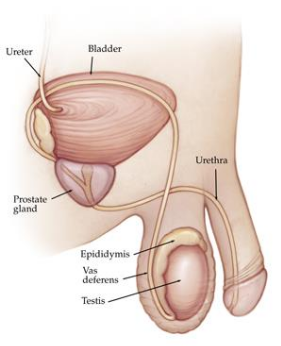
First of all, the prostate is under the bladder, shaped like the pointed one of chestnut faces towards the penis, and the urethra is lodged in the interior leading to the urethra in the penis. The function of the prostate secretes the liquid component of semen and helps sperm movement.


1
The prostate is an organ only in men, it does not exist in women. It is embryologically equivalent to a woman's uterus. Cancer that occurs in this prostate is called prostate cancer.

Epidemiology
I will show male cancer incidence in Japan and future prospects.
2
I can see that prostate cancer has increased markedly. In 2020, it is estimated that it will be the second largest affected number after lung cancer. The background of the increase is related to the fact that as society ages, the dietary habits become westernized, and the diagnosis technology advances, many things have come to be discovered.
I will show you the incidence of prostate cancer by age.
It turns out that prostate cancer is a typical elderly cancer that increases with age.

Diagnosis
As a test and diagnosis of prostate cancer, we conduct tumor marker, PSA examination, rectal examination, ultrasound examination by blood test as examination to find a person with cancer possibility. If you suspect cancer here, you will do a prostate biopsy and tissue examination to confirm cancer, but before you undergo a biopsy to see where in the prostate cancer is likely to occur, a tomogram called MRI I will take. When cancer is confirmed, we will examine bone metastasis called CT tomography and bone scintigraphy to investigate the spread of cancer.

3
PSA test is a convenient method of finding prostate cancer by blood test. "Prostate" "Unique" "Antigen" English initial letters were attached. PSA is a type of protein made in the prostate and exists in the blood even when it is healthy but it is used as a tumor marker for prostate cancer because a large amount of PSA flows out into the blood when prostate cancer develops It began.

4.
The relationship between PSA value and prostate cancer detection rate, the higher the PSA value, the higher the likelihood of prostate cancer. If the PSA value is higher than 4, prostate cancer is suspected, 10 in 1/3 and 1/20 in half, and in more than 50 in prostate cancer is found in most people. Also, the possibility of metastasis will also come if you get 50 or more.

5.
If prostate cancer is suspected, a tissue examination called prostate biopsy will be conducted, but under anesthesia, ultrasound is inserted from the anus, a fine needle is stabbed while observing the image, about 10 prostate tissues are collected To do. Then examine with a microscope for cancer. Because I do anesthesia, there is hardly any pain.

6
Although it is an image examination, in MRI, it is investigated whether there is spread to the cancer place and surroundings, in CT, whether the lymph nodes and other organs have metastasis.

7
Bone scintigraph examines the presence or absence of bone metastasis. The place where the transition exists is blackened. With X-rays, the place where the transition exists is whiter than other places.

8
Treatment
Treatment for prostate cancer can be broadly divided into treatment to the whole body and treatment to the prostate. There is hormonal therapy for the treatment of the whole body, but there are treatments of medication treatment and removal of the testicles. Treatment for the prostate, the surgical treatment will remove all the prostate. As radiation treatment, there are methods of irradiating from outside the body and irradiation from the inside of the body.

9
Hormonal therapy
Male hormones promote proliferation of cancer cells in the prostate. Hormonal therapy is the reverse of this. Prostate cancer growth is suppressed and the cancer is reduced as a result of removing the testicles or by taking medicine to prevent the transition of the nutritional source, androgen, to prostate cancer. In medicine, there are drinking drugs that suppress the actions of male hormones, and injection medicines that suppress the secretion of male hormones. If the effect of hormonal therapy is lost, treatment of anticancer drugs is necessary.


Surgical treatment
Surgery is called total prostatectomy. Laparotomy was a standard procedure in the operation to remove the seminal vesicle attached with the prostate, and gradually moved to laparoscopic surgery


10
Laparoscopic surgery is a surgical operation performed by observing the endoscope image with inflating carbon dioxide and carbon dioxide in the stomach like this. Insert the forceps and the endoscope with the instrument called a trocar as a passage

11
In laparoscopic surgery, the surgeon stands on the patient's left side, assistant, photographer on the right side. Then surgery is performed while watching the monitor on the foot side

12
It is the first advantage of laparoscopic surgery for prostate cancer, but it is not only small and inconspicuous but also has a mild postoperative pain and rehabilitation faster. Although it is surgical wound, laparotomy surgery enters about 20 cm incision in the lower abdomen, but in laparoscopy there are only 5 small scratches from 5 to 12 mm

13
The second advantage is that the pressure due to pneumoperitoneum results in a very small amount of bleeding compared to laparotomy, requiring transfusion is rare. As a third advantage, because of the enlarged visual field through the endoscope, it is expected to improve cancer cancers and postoperative quality of life (urinary incontinence, deterioration of sexual function) by delicate surgery. The probability of resection is the probability of cancer at the end of the resected prostate, but in our hospital it is also lower than laparotomy with laparoscopic surgery, the rate of recurrence is small and laparoscopic surgery is radical It is clear that it is excellent in the ability to cure cancer. As for surgical operation time, it is technically difficult compared to laparotomy surgery, so it is said that surgery is long, but our hospital has no difference from laparotomy with 2 hours to 2 and a half hours (median 2 hours 25 minutes) .


Radiation treatment
It is a treatment method which irradiates radiation and kills cancer cells. Since the burden on the body is less than surgery, treatment is possible for elderly people and people who have heavy medical history. There are two methods of irradiation from the outside of the body and irradiation from inside the body. A therapy that radiates radiation to the prostate from outside the body keeps a mark on the skin and irradiates the radiation there. One irradiation time is 5 minutes, there is no pain due to irradiation. Therefore, even outpatient treatment is possible. Usually, I continue to irradiate 5 days a week once a day for 7 weeks. Inflammation to the place you see is seen as a complication during treatment. If inflammation of the skin or inflammation of the bladder occurs, urination pain and frequent urine are seen, and diarrhea and anal pain are seen when the rectum is inflamed. There is bleeding as a complication of late period for a while after treatment, hematuria and blood stool may be seen. However, due to the progress of instruments, complications are decreasing considerably. Irradiation from the body is a new treatment that embeds the source of radiation inside the prostate and kills cancer cells. Insert the source of radiation while watching with ultrasound. The time is about a few hours. It is a short period, but hospitalization is necessary. If you have cancer at a very early stage, you can expect the same therapeutic effect as surgery.
7/21/2018 01:00:00 PM

Today is the day to think about eating.

Eating out inevitably tends to get frustrated in hurried days. I took breakfast, I did not eat much vegetables. The reason why we can not maintain a good balance is "Is there anything else you care about and there is not enough room to think about that!" Is it honest?

Nonetheless, I think that I think that I can try a healthy meal today.

On April 19th "Food Day Day"


First of all, 19 days of each month by the Cabinet Office the day of the food education , but I have been defined as (※ 1) . In addition, on April 19, it seems that it is set as the day of food education by private companies (* 2) . In other words, it is the day of double food education .


(* 1) Established in 2005 according to the basic education promotion promotion plan based on the Basic Eating Law.
(* 2) Mitsubishi Corporation established from a pun of 419. The enactment date is unknown.

A little more healthy meal than usual


I do not say difficult things to eat according to the " meal balance guide " that I learned in lessons when I was a junior high or high school student . I just want you to try something more healthy everyday than usual.
7/21/2018 12:58:00 PM

Four points to promote "rice" from a controlled nutritionist "bread"

Regardless of age and gender, breakfast has many bread schools. However, it is often said that staple food is healthier and better than old. What is it in the first place?


If you look only at calories, there should not be much difference. Rather, it has a lower calorie than a bowl of rice bowl (1 cup of rice: 269 kcal, bread 1/6 cuts: 158 kcal) compared to bread.

Mr. Kazuki Enone of a managed nutritionist says, "At the end of the day it is simple but at the end of the day to eat" (wanibooks), from these four points, we prefer "rice" rather than "bread" to staple food It is talking. Even so, I can not stop the bread habit in the morning (laugh)

4871218427592704
© aluxum / iStock
01.
Lipid: rice <Bread
When you compare 1 cup of rice with bread (6 slices), the calorie is indeed lower in bread. However, lipid, rice: About 0.5 g, (depending on the type) bread: about 2 - 4 g is a standard. It contains several times as many lipids as rice .

02.
No rice is added
The biggest difference is this. If you just cook white rice, no rice is added. On the other hand, bread has oil, sugar, salt and so on, and many contain additives such as yeast food, emulsifier, Na acetate, sweetener, flavor and so on. Incidentally, "trans fatty acid" which is sometimes included in bread is a component that becomes subject to regulation in the whole country as it is harmful to the body.

03.
Side dishes calories and lipids
There are many low-calorie Japanese dishes such as grilled fish, miso soup, simmered dish, and natto, which are often combined with rice. In the case of bread, those with high lipids such as bacon egg and potage soup match well. If you apply jam or butter, it is natural that it will be more calorie.

04.
Number of bites
Even with a stubbornness, it is ritual to rice. As with rice, grain eating increases the frequency of chewing, making it easier to get a feeling of fullness. It is also easy to prevent snacking if you do so.

...... But I like bread!
From this point on, rice is still healthier. But I can not stop tasty bread! It is ideal that the ratio of main meal of rice and other food (including noodles) is about 9: 1. If you eat 3 meals every day , we recommend a balance of about one meal on the 3rd .

By the way, if you eat bread, the one with low degree of whitening such as whole grains is healthier.
7/21/2018 12:56:00 PM

To people who want to start running. Let's hold down only these 5 points before running.

I want to exercise for the summer! Running seems to be the easiest, even if you do not join the gym. But, I'm not good at driving ... .... The model of Victoria Secret, Karlie Kloss, was also originally disliked running. Such she is Caroline Burke's that was conscious in order to finish the full marathon and sunny the " Elite Daily introduced me to in the article of".


Let's start with this as a trigger !
6365347724656640
For running, in particular for inexperienced people, the hurdles are expensive and I do not know how to approach them. It may be the workout method of Karlie Kloss who will give you such a heavy back .

She started running as a training for the half marathon in Paris and finished the first full marathon in New York as soon as possible . Now is also the same time entrepreneurs and there under the name model , the cover of "Forbes" celebrities decorate up.

Even if I can not imagine myself who is still running now, I would like to take advice from her seriously. I will introduce "5 aims" that will lead you to "runner's high" with Kloss's comments.

01.
Aim for
sustainability by finding the environment to be inspired
5529879948820480
Each person has different circumstances in which results can be obtained. Then, let's first find an "environment where you can enjoy running". Some people like to run with running machines that will instantly run speed and distance, others who like outdoor running that throws themselves naturally.

Karlie Kloss is a sudden outdoor faction . She talks like this.

I do not seem to be suitable for running machines. I think it took time to start running, because I was trying my machine all the time. The scenery does not change, it does not fit in imaichi ne.

02.
I think to succeed
Movement of the body may be "physical activity" to the last, but it is also fact that the mental state greatly affects it. In other words, to maintain motivation for workouts, it is important to have a mindset of "to succeed". Until you can do it, you only need to do "pretending", so it is key point to challenge with confidence.

Kloss talks about the rejection that he had in running .

At that time, I was deciding my own thing. "I am not a runner". But when I cease to decide, I finally got to think "Let's challenge the half marathon, let's run."

03.
Have a certain level of competitiveness
5851299195125760
Just having a little competitive spirit, you have to follow the next step.

Of course, getting competitive spirit and support from running companions is also a hand, but myself is a sufficient competitor. For example, to run faster than last time, aim for a long distance. It is a wonderful ambition to aim both of them.

Kloss also implied the importance of competitiveness .

I am originally competitive. So when there are people who can run faster than I can, I'll push myself as closely as possible to match them.

04. Carefully routine
after workout
The quality of the workout is dependent on whether you regularly work from your body. Therefore, let's try not to lose your stretch, hydration and then ingest the necessary nutrition so that you can fully recover after running.

Advice for Kloss to deliver voices against running is to take moisture firmly.

Anyway to drink plenty of water! I try to take plenty of water before and after workout. Afterwards I'm charged with energy by drinking protein shake and ingesting the necessary nutrition.
05. Grasp
running as a change of pace
5016717256294400
Running is a good opportunity to rest your brain one step away from a chaotic and busy day and to check your own axis. Some people refresh their minds with meditation, but just by jogging with sneakers can make the brain clearer .

The following is from Kloss' s interview.

Running is the time you can look back on yourself without being disturbed by anything. So I feel like refreshing. Because you can bring only what is really important to you to the axis of your mind, after running, you can think "OK, I will do my best tomorrow."
7/21/2018 12:12:00 PM

Colorectal cancer

Introduction
Cancer has become a very familiar illness. In life, one in two people will be cancer, one in three will die from cancer. It is a colorectal cancer that is one of the five major cancers, but it is said that 100,000 people are going to have colon cancer every year, aging and eating habits to Westernized, these two points increase colon cancer It is related.
The wave of aging is difficult to do anything, and what we can do to eradicate colorectal cancer is in the early detection by preventive and medical examination including improvement of dietary habits. However, there is a limit to "Taking one or two foods and nutrients in the diet into issues" in order to prevent specific cancers.
We recommend balanced diet and abdomen, moderate exercise, a good night's sleep, participation in medical examinations. Below is an overview of colorectal cancer.

Diagnosis of colorectal cancer
The length of the colon in general adults is said to be about 1.5 m. Symptoms vary slightly depending on where you can do the same colorectal cancer. For example, if it is on the right side of the large intestine, it touches anemia and a lump, on the left side it is abdomen, constipation finally is bowel obstruction, and furthermore when approaching to the anus side blood mixes with feces (Fig. 1). If you can do it, I would like to have a diagnosis before such symptoms appear.

For early diagnosis
There is a fecal occult blood test that senses a small amount of blood contained in feces from colon cancer and is used for colon cancer screening. If fecal occult blood was positive (a little blood is contained in the stool), further examination is required. However, fecal occult blood is not equal colorectal cancer. In cases where it becomes positive with colon polyp, enteritis, hemorrhoid disease, etc., and, as a result, the cause is not clear, is the majority.
On the other hand, fecal occult blood negatives are not safe. It is because it is not always bleeding from colorectal cancer. PET examination is also a cumbersome and too expensive examination method to find out early colorectal cancer from a large number of people efficiently.
Unfortunately there is no simple, cheap and reliable diagnostic method leading to diagnosis at an early stage. It is a practical way to consult a doctor if you are aware of the symptoms mentioned above or you have a large number of cancer patients in families / relatives with blood, while regularly taking advantage of medical examinations and human dogs.

To diagnose the presence of colorectal cancer and its part
After colonoscopy examining the large intestine by inserting an endoscope of about 1.3 m in length from the anus, we undergo CT colonography at our hospital. It is an alternative to the colonoscopy examination which indirectly looks inside the large intestine by popping barium and air from the anus which had been done so far and it became possible to visualize the running of the blood vessel simultaneously (Fig. 2, 3). 

Once diagnosed as colon cancer
If necessary, check whether you are stuck (invading) to the organs in the vicinity of the colon cancer, whether it is flying (transiting to distant organs), chest X-ray examination, CT examination or abdominal echocardiography We will investigate in detail.

Treatment of colorectal cancer
From the point of view of treatment
There are 4 types of surgical therapy (including endoscopic treatment), chemotherapy, radiotherapy, and others.
Laparoscopic surgery: Depending on the progress and size of the cancer, surgery for inserting a television camera or surgical instrument from a tiny wound with tummy is performed actively in our hospital.

From the viewpoint of therapeutic purpose
There are 4 kinds of treatment for cancer which becomes a primary cause in the large intestine, treatment for metastasis, treatment for preventing recurrence.
If the spread of cancer is limited, surgical therapy and radiation therapy are performed, and when cancer is spread throughout the body, chemotherapy is performed. Immunotherapy and gene therapy are still not common.

What is adjuvant therapy
Needless to say, even in the case of early cancer, there are cases where cancer spreads along the flow of blood and lymph at a site away from the cancer lesion that is the primary cause of cancer. After surgery sufficiently removes the cancer, the treatment to control such spread is called postoperative adjuvant therapy. Its purpose is to prevent recurrence, and chemotherapy and radiation therapy can be cited as a treatment method.
On the other hand, there is also a preoperative adjuvant therapy. The aim is to reduce cancer by performing chemotherapy or radiotherapy before surgery for highly advanced cancer. Those that could not be removed become resectable, and in addition, it may be possible to resect with small surgery.

About chemotherapy
Chemotherapy is required depending on the degree of cancer progression (stage). The drugs used include anticancer drugs that kill cancer cells, 5-FU (5-FU), irinotecan (CPT-11) developed in Japan, oxaliplatin, trifluridine, and further cancer Bevacizumab, which is an angiogenesis inhibitor (which is an angiogenesis inhibitor), cetuximab and panitumumab, which is an anti-EGFR antibody that blocks part of the network of cancer cells, and molecular targeted drugs such as legorafenib that blocks in a wider range.

Internal treatment
Treatment by oral administration is also reviewed.
Oral drugs of 5-FU type developed in Japan are attracting attention in Europe and the United States. As adjuvant chemotherapy for postoperative adjuvant chemotherapy for colorectal cancer that has progressed to a certain extent, it is simple compared to intravenous injection and the outcome of treatment is comparable. Even in the case of highly advanced cancer or recurrence which is difficult to resect by surgery, by combining with injectable medicine, a treatment method comparable to the outcome of treatment can be compared with the method by only injection, It came to be seen.

Exogenous chemotherapy
In our hospital considering the patient's burden, we are focusing on outpatient chemotherapy. In chemotherapy combined with the above-mentioned molecular targeted drugs, annual expenses may exceed 10 million yen, so there is a high medical care cost system to alleviate individual burden.

On side effects of antineoplastic drugs
Anticancer drugs are taken up more frequently in cell proliferation rate. Generally cancer cells are expected to be effective because cell division proliferation is active. However, normal human body also has a population of divisive proliferation. Stem cells in the bone marrow that make the origin of blood (blood cells), gastrointestinal tract, reproductive system, cells in the root of the hair. Naturally, many of anticancer drugs are taken into these cells and will be affected. Therefore, side effects such as myelosuppression, nausea / vomiting, diarrhea, hair loss etc. will appear. Although it is called a side effect, it can be said to be the original action if considering the property of anticancer drug that it is taken in and acts on cells with fast growth rate.
However, when chemotherapy is completed, many side effects will be eliminated promptly. It may take more than a few months to completely recover. On the other hand, there is an individual difference. Some race, or even the same Japanese people, have side effects strongly. Irinotecan (CPT - 11) is a commonly used anticancer agent for colorectal cancer, but in people with abnormal enzyme related to this there is a strong toxicity of (CPT - 11). A certain degree of discrimination is possible by genetic testing.
On the other hand, it is a molecular target drug, but there are special side effects. Cancer will make a new blood vessel to secure necessary blood to grow cancer, but there is medicine called bevacizumab (released in June 2007) to suppress this. Although side effects include hypertension and nose bleeding, serious things such as arterial occlusion are rarely caused.
We exchange information to proliferate cancer cells. Cetuximab (released in July 2008) and panitumumab (released in June 2010) are anti-EGFR antibodies that block a part of this information network. Adverse events are mainly skin disorders such as rashes and skin cracks. From April 2010, K-RAS gene mutation testing, which determines the therapeutic effect beforehand, is now undertaken in insurance medical treatment. In May, 20th, Legolafenib which blocks a wider range of information networks was released, there are many skin disorders as side effects, high blood pressure, bleeding, rarely liver failure, etc.

Friday, July 20, 2018

7/20/2018 11:59:00 PM

Origin and treatment of hepatocellular carcinoma (HCC)

Ⅰ. Introduction
The number of deaths of primary liver cancer in our country increased from 8291 (1958) to 34637 (2002). By gender, men were 23,815 (2002), women was 11822 (2002). After that, the number of deceased people has decreased, but it exceeds 30,000 a year in the past.
Possible cause of decrease in the number of liver cancer deaths are as follows:
1) Increase in other diseases and deaths due to the aging of the age group of primary liver cancer
2) Diffusion of periodic imaging diagnosis for hepatitis virus positive people accounting for approximately 90% of patients Increase in early detection cases by
③ Increase in carcinogenesis by antiviral therapy
④ Advances in the treatment of liver cancer


may be considered. 
However, because hepatocellular carcinoma is poor in subjective symptoms, it is still found in advanced hepatocellular carcinoma, except for patients who are enclosed as high-risk group of liver cancer and regular imaging diagnosis is done Current situation is that there are many cases.
Progressive hepatocellular carcinoma is not clearly defined, but (
1) giant liver cancer with a maximum tumor size greater than 10 cm (
2) cases with advanced vascular invasion (
3) multiple cases in the liver (
4)
a case with distant metastasis is advanced In addition, it is difficult to treat patients with hepatopathy degree C or Child-PughC in which the liver reserve capacity has decreased. For each case, it is a principle to accurately grasp the pathology such as primary tumor, vascular invasion, metastatic lesion, liver reserve and the like, and to perform custom-made treatment according to each case in principle, and a multidisciplinary treatment is desired I will.

Ⅱ. Origin of hepatocellular carcinoma
Cirrhosis and liver cancer in Asian countries are dominated by HBV-positive individuals. For example, in Korea, 74% of liver cancer is positive for HBs antigen, HCV antibody positivity is 9%, alcoholicity is only 7%, while the cause of cirrhosis and liver cancer in our country is few HBV and HCV positive It is occupied principally. At the 44 th Annual Meeting of the Japanese Society of Hepatology held in 2008, results of each institution were gathered from the main facilities nationwide, and the causes were analyzed for 17262 cases of liver cirrhosis (FIG. 1) and 16117 cases of hepatocellular carcinoma (FIG. 2) It was. In Japan overall, 73% of liver cancer is positive for HCV antibody, 14% is positive for HBs antigen, and it is very different from South Korea's achievement. In Japan, the cause of cirrhosis of cirrhosis has been investigated 5 times from the General Assembly of the Japanese Gastroenterology Society of 1983 to the Japan Society of Hepatic Societies in 2011. B type hepatocellular carcinoma is almost constant in the number of deceased but due to the increase in type C hepatocellular carcinoma the proportion of type B has decreased from 23.3% in 1983 to 13.1% in 2011 I will. In addition, in the results after 1998, which is a survey after the diagnosis of HCV was established, the non-B non-C type increased from 21.8% to 26.2%. It is believed that both B and C hepatocellular carcinomas will decrease in the future and non-B non-C hepatocellular carcinomas such as NASH (nonalcoholic steatohepatitis) liver cancer will increase.

Ⅲ. Progression classification of hepatocellular carcinoma and treatment algorithm
Progression classification and prognosis
The degree of progression of hepatocarcinoma described in the handling regulation of primary liver cancer is classified as Stage I to IV according to the highest numerical value of each patient by obtaining the progress value of that patient by each item. Life prognosis of hepatocellular carcinoma greatly influences not only tumor progression but also liver reserve. In the treatment regulations for primary liver cancer, we present classification of hepatopathy degree incorporating ICG burden test instead of hepatic encephalopathy in Child-Pugh classification. The 5-year survival rate of hepatectomized cases was 59%, 45.3%, and 35.0% for liver damage degree A, B, and C, respectively.

Ⅴ. in conclusion
Even at our hospital, any treatment other than liver transplantation is possible, but it is important to start treatment at the stage of chronic hepatitis because it is not cancer. For hepatitis C, it changed from traditional interferon treatment to treatment with oral medicine, and more than 90% virus removal became possible. Also in hepatitis B it has become possible to stabilize hepatitis and prevention of cancer with nucleic acid analog formulation. Early diagnosis of NASH (nonalcoholic steatohepatitis), which is expected to increase in the future, will lead to the prevention of NASH liver cancer.
7/20/2018 11:57:00 PM

About stomach cancer

Introduction
Currently, malignant neoplasms are the No. 1 mortality and mortality by type of death cause, malignant neoplasms are death by about 1 in 3.5 people. Moreover, it is increasing trend year by year. Looking at malignant neoplasms by organs, gastric cancer is the second largest in both males and females. In other words, gastric cancer is not a special disease, it can be said that it can happen to anyone.
First of all, what we can do is to grasp and prevent the risk factors of gastric cancer and to discover it at a time when cure is possible.


What is stomach cancer
The wall of the stomach is divided into several layers. Stomach cancer is a cell of its innermost layer (mucous membranes secreting gastric juice and mucus) has been mutated to various cancer cells as a result of various factors. Also, stomach cancers are classified into several types by inspection with a microscope. Even with the same stomach cancer, personality is different little by little depending on its type, there are differences in treatment method · treatment effect etc.

Causes of stomach cancer
At present, reliable risk factors include smoking and H. Pylori infection. Especially for H. Pylori infection, the risk of cancer is 5.1 times higher than non-infected persons. Besides, taking too much salt, diabetes etc as a risk factor is said to be said. Conversely, green tea, vegetables and fruits are considered as inhibitors of gastric cancer.
* Currently, for H. Pylori infection, eradication therapy is insurance indication. The risk of developing gastric cancer will be reduced by sterilization (it can not completely suppress gastric cancer).

Symptoms of stomach cancer
The main symptoms are stomach pain / stomach discomfort, heartburn, nausea, loss of appetite, frequent occurrence of belching etc. However, neither symptom is unique symptoms of stomach cancer. In addition, it is asymptomatic in most cases at early stages, and it may be asymptomatic even if it progresses.

Diagnosis
Firstly, if any symptoms are recognized, please consult a medical institution earlier.
Various inspections are selected as necessary. Currently, when it is judged that there is a possibility of gastric cancer, in many cases upper endoscopy is chosen. If it is diagnosed as stomach cancer (including suspicion) by endoscopic examination, a cancer tissue is collected and a tissue examination (examination by a microscope) is carried out to make definite diagnosis as stomach cancer. When the diagnosis of stomach cancer is confirmed, investigate the presence or absence of metastasis by conducting contrast CT or PET-CT examination to determine treatment policy.

Treatment
The treatment of stomach cancer is determined based on the degree of progress (stage, stage) of cancer. Currently, there are three focused on endoscopic therapy, surgical therapy, chemotherapy (anticancer drug treatment).At present, gastric cancer is found to be a curable disease if it is found early and appropriate treatment is done. In addition to striving to prevent gastric cancer, it is important to undergo periodic examinations, and to consult a medical institute as soon as you feel any discomfort or symptoms.

Efforts about stomach cancer at our hospital
Our hospital proactively recommends endoscopic examination for patients who have been consulted with complaints of discomfort in the stomach. As a result, there are cases that lead to the discovery of ultra early cancer. Also, depending on the stage, we are actively working on endoscopic treatment as well.
Currently, we regularly hold a study group on diagnosis / treatment selection and treatment outcome for each case, mainly of gastroenterology internal medicine, surgery and clinical pathology science, and are working together in mutual cooperation.